
Man does not become human,
but is always human.
Erich Blechschmidt


The third week: the embryo gets direction
In the second week the embryo grew explosively in the periphery. The tissue between the outer trophoblast and the inner embryoblast tore away and resulted in the chorion cavity. If the process of strong growth in the periphery and little growth inside would continue, a growing and ultimately unbridgeable distance would arise between the nutritive trophoblast and the embryonic disc. To stop that process a reversal takes place in the third week. The periphery will develop further, but the focus of the development will now be on the embryonic disc.
Several developments occur more or less simultaneously. For easy reference, they will be discussed separately in the order in which they occur.
Gastrulation: the formation of mesoderm tissue
The first process that takes place in the third week is called gastrulation (gaster = stomach). For simple animals gastrulation means the formation of a stomach and a digestive tract (see the page: plant and animal). At the same time the body cavity is formed in which the mesoderm develops. For humans, gastrulation is the process whereby only the mesoderm is formed. The digestive tract develops later in a process in which the amnion is involved.
At the beginning of the third week the tissue of the epiblast thickens and a small indentation is formed in the embryonic disc near the connective stalk. This occurs at the caudal end of the embryonic disc (cauda = tail, caudal = at the tail end; Fig. 20 and 21A). This formation is called the primitive streak. The primitive streak grows in cranial direction (= side of the head), i.e. away from the connective stalk. From the primitive streak mesoderm cells are ligated, which migrate to the space between the epiblast and the hypoblast. In this process the cells of the hypoblast are replaced by these newly formed cells, too.

Figure 20. Gastrulation in humans (similar to Fig. 21-A1)
Cross-section through the embryonic disc in which the ligation of mesoderm cells from the primitive streak in the epiblast (cubic cells) is visible. The mesoderm cells migrate to the space between the epiblast and the hypoblast and replace the flat cells of the hypoblast, too.
The formation of the notochord is here only given in outlines. The actual process is more complicated. See: www.embryology.ch
The primitive streak grows to the middle of the embryo (Fig. 21B). There it makes the primitive knot. This is the place where most mesoderm cells are ligated. These cells migrate to any place between the epi- and hypoblast, except for the prochordale plate and the places where the mouth and anus will arise, because epi- and hypoblast stick together at these places. The epiblast is now called ectoderm and the hypoblast entoderm.
Then a tube is formed that grows from the primitive knot between the epi- and hypoblast to the prochordale plate. This tube is called the notochord (= chorda dorsalis; Fig. 21C). This flexible rod gives the embryonic disc solidity and creates a symmetry-axis in the embryo. The notochord also forms the directions left and right; the mesoderm gives the embryonic disc content and thickness. The notochord is the formation around which the vertebrae will grow. In an adult small remnants are visible in the intervertebral discs.
At the same time, the embryo grows rapidly in length and not so much in width, and faster at the cranial side than at the caudal side. Thereby the form changes from round to oval. Because the embryo grows faster at the cranial end, the importance of the primitive streak diminishes and it disappears eventually.

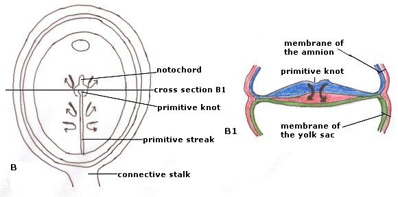
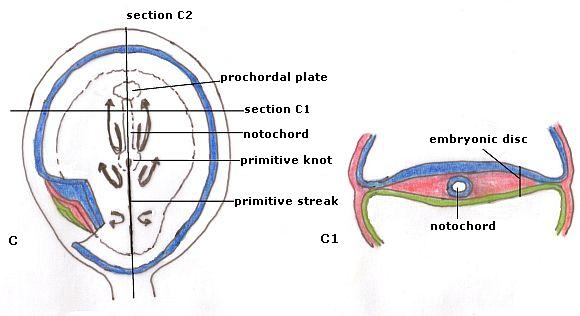
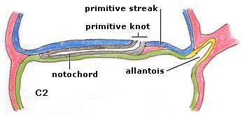
Figure 21. Gastrulation or the formation of the mesoderm (simplified)
A, B and C Dorsal view
A1, B1 and C1 are the corresponding cross-sections C2 is a longitudinal section of C
A: at the caudal end the primitive streak grows toward the centre of the embryonic disc. From the ectoderm mesoderm cells are ligated to the space between the epi- and the hypoblast.
B: In the middle of the embryonic disc at the end of the primitive streak arises the primitive knot. This is the place where most mesoderm cells arise, that migrate to all parts between the ecto- and the entoderm. From the primitive knot the notochord is created, a tube that runs between ecto- and entoderm.
C: The notochord runs until the prochordale plate. Between ecto- and entoderm is everywhere mesoderm.
C2: a longitudinal section showing how the notochord runs. At the same time as the notochord the allantois is formed. This is a protrusion from
the yolk sac into the connective stalk.
The neural tube and the somites
The genesis of the neural tube is related to the growth and development of the notochord. From the 19th day, the ectoderm above the notochord gets thicker (Fig. 22). This formation, about half of the ectoderm, is called the neural plate. The central nervous system will develop from this tissue. The rest of the ectoderm does not thicken and will become the skin.
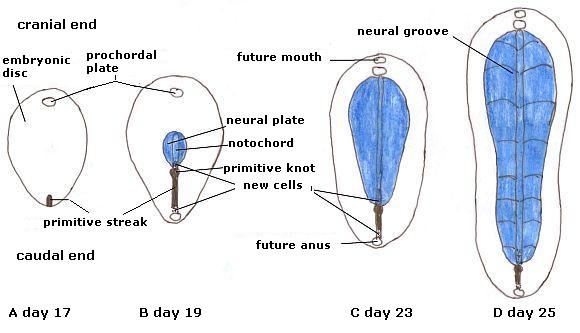
Figure 22. Dorsal view of the embryonic disc. Development of the notochord and neural plate (dating and interpretation prochordale plate: www.embryology.ch)
Only ectoderm is shown, the uncoloured tissue would be blue, too. For clarity, only the neural
plate is coloured. The uncoloured ectoderm becomes the skin.
A. On day 17 caudally the primitive streak appears, that grows to the centre of the embryo and ends in the primitive knot.
B. Here one can see that the notochord grows from primitive knot to the prochordale plate. Above the notochord the ectoderm tissue thickens and constitutes the neural plate. The primitive streak and notochord grow from the caudal to the cranial side.
In C. the embryonic disc becomes elongated as growth occurs mainly at the cranial side. The notochord has grown until the prochordale plate.
D. The neural plate folds itself from the middle into the neural groove. The primitive streak barely grows, the notochord does.
The embryonic disc is growing fast: in five days it doubles its length. On the cranial side it grows much faster than on the caudal side. This makes the neural plate at the head longer and wider than at the side of the tail. The thick neural plate (Fig. 23 and 24) bends in, first in the middle and from there to the cranial and caudal ends. The indentation becomes deeper and the neural groove is formed (Fig. 23 and 24). The connection with the ectoderm is broken and the neural groove and the ectoderm close. The neural tube is created. From the middle of the embryonic disc, this process goes to both ends. The neural tube closes at the cranial end on the 29th day, one day earlier than at the caudal end (30th day).
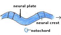
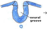


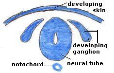
Figure 23. The development of the neural tube (cross-sections)
The thickened ectoderm bends into the neural groove. At the ends is the neural crest. The neural groove deepens and the neural crest expands (third image). On the second row the neural tube is visible. From the neural crest ganglia arise. The neural tube is located between the skin and the notochord and will develop into the spinal cord.
In the neural tube there is amniotic fluid. From the neural crest ganglia will arise, that lie next to the spinal cord and spread out over the body. Left and right of the neural tube paired clusters of mesoderm cells develop, called somites (soma = body; Fig. 24), about 40 pairs in total. The vertebrae and muscles will eventually develop from these.
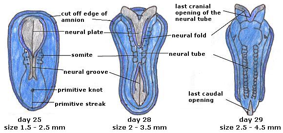
Figure 24. The development of the neural tube; dorsal view (www.embryology.ch). All tissue is ectoderm, the neural plate is coloured gray.
From left to right days 25, 28 and 29.
The closure of the neural tube begins in the middle. Next to the neural tube the somites develop, they are visible as bulges under the ectoderm.
The embryonic disc grows from about 2 to 3.5 mm in five days.
The allantois
At the beginning of the third week the yolk sac grows out finger-like into the connective stalk (Fig. 21 C2). This small organ is called the allantois (allantos = sausage). It plays a role in the development of blood and the circulatory system. Later it participates in the formation of the bladder. In humans it remains small, in embryonic birds the allantois is the organ for breathing.
The blood and its circulation
In the second week, the embryonic disc is so small that nutrition and oxygen from the mother can reach the cells by diffusion in the intercellular liquid. Disposal of waste products takes place in the same way. The growth of the embryonic disc in the third week causes this to be no longer sufficient and a new transport-system is needed. This is created from the middle of the third week on.
At first, the blood vessels and the blood develop in the mesoderm on the outside of the yolk sac and the allantois. There, clusters of cells are formed, called blood islands (Fig. 25). In the blood islands cavities arise. The blood islands grow and several blood islands connect with each other. Then the cavities merge, so that capillaries are formed. The capillaries become longer and find their way to the embryonic disc. Some cells in the wall are transformed into blood cells that will flow in the liquid in the capillaries.
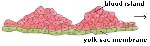
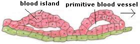
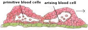
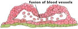
Figure 25. The development of blood vessels
First clusters of cells arise, blood islands, in which cavities occur. These cavities grow together and capillaries arise. From the same primitive tissue cells blood cells emerge simultaneously, which will flow in the stream of liquid.
Blood is first formed outside the embryonic disc. In the embryo itself it is not formed until the fifth week.
The blood starts to flow because in the embryonic disc some substances are required and others are expelled. The blood flows in capillaries in the embryo on the ventral side towards the head, where its flow is stopped by firmer tissue. The blood must turn around to be able to flow back on the dorsal side of the embryonic disc to the connective stalk and the trophoblast. In this movement, congestion occurs and the stream of blood is stopped for a short while. This is why the blood starts to pulsate. In this place (i.e. on the head side of the neural tube; above the tissues that will become the head) the heart is created (Fig. 26). Around the 20th day it starts to beat.

Figure 26. Position of the heart and the circulation at the end of the third week
The heart lies cranially of the neural tissue and the mouth-membrane (the thin spot behind the heart). The blood flows ventrally (on the side of the stomach) to the head and dorsally (the back side) back to the connective stalk (the arrows are drawn outside the embryonic disc, however, the blood flows in it).
In the beginning we see a flow of liquid, and because the stream meets with firmer tissue, causing congestion and stagnation, the beating heart comes into being. This phenomenon is also seen when a stream collides with a hard material, like waves breaking on the coast, water meeting stones in streams and flow-forms.
The heart arises from flow and stagnation, it is not the cause of the flow of blood. The heart is the only place where the flowing blood comes to a standstill for a short time.
The circulatory system is the first organ in operation.
The chorion and the trophoblast
The rampant growth of the trophoblast decreases towards the end of the third week. The syncytiotrophoblast develops cell membranes and will now be called cytotrophoblast. It constitutes the binding layer between the chorion and the uterine tissue. Blood vessels develop in the chorion which run from the connective stalk to the villi (= flakes), where the exchange of substances with the blood of the mother takes place from the end of this week.
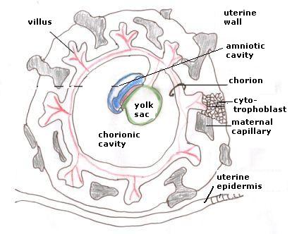
Figure 27. The development of the chorion and the placenta
The embryonic disc with amnion and yolk sac is attached to the connective stalk in the chorionic cavity. In the chorion the blood vessels are indicated that run from the connective stalk to the villi. The syncytiotrophoblast has become cell membranes and is now called cytotrofoblast.
Characteristics
The focus of the development of the embryo in the third week is on the embryonic disc. There is still much growth in the periphery, but it is no longer a proliferating growth and the tissue gets cell membranes. There is now a relative calmness in the periphery. If the growth-movement of the second week would continue, man would stay in the periphery (or his enclosing organs) and would not develop a body.
From the periphery (yolk sac, connective stalk) blood circulation arises, finding its centre in the heart. The heart arises from the flow of blood. Pulsation of the heart arises from standstill and flow.
Simultaneously, thickness is created in the embryonic disc by the ligation of mesoderm cells from the primitive streak and -knot. This gives the embryonic disc content. Left and right are created by the formation of the notochord, later reinforced by the formation of the neural tube. The embryonic disc has become a spatial, three-dimensional structure.
An important point in the embryonic development of man is around the 17th day, when circulation and heart are created. If not, the development cannot go any further and stops.