
Man does not become human,
but is always human.
Erich Blechschmidt


The second week: growing out in a new shelter
This week the embryo nestles in the wall of the uterus. The tissue on the periphery, the nutritive trophoblast, grows fast. From the inner cell mass, the embryoblast, the embryonic disc will be formed. The embryonic disc becomes largely disconnected from the trophoblast. Differences in growthrate are responsible for this process.
Implantation
The embryo (the blastula) is out of its rigid zona pellucida and has arrived in the uterus. It can now grow and it implants itself on the side of the embryoblast into the wall of the uterus. Enzymes of the embryo digest the maternal uterus tissue. The embryo invades; it eats into the wall of the uterus. The embryo behaves aggressively.
The trophoblast grows fast, so fast that it causes a proliferative tissue with many nuclei and without cell membranes (called syncytiotrophoblast (syn = together, cyto = cell). A layer of 'normal' (cyto)trophoblast cells (= nutritive tissue with cell membranes) remains present between the syncytiotrophoblast and the embryoblast.
In the syncytiotrophoblast gaps arise, called lacunae, through which maternal blood starts to flow. Only one membrane exists between maternal blood and embryonic tissue, and there is just one barrier for the exchange of substances. Embryonic tissue also surrounds capillaries and maternal glands. In this way, the embryo can be supplied with oxygen and nutrients and waste products can be disposed of. However, substances that are bad for the embryo can also get through.
The embryo eats into the maternal tissue. On the other hand, the mother gives room to the embryo in her own tissue. She allows a strange creature to grow in her own body. This is a wonderful process, because strange creatures (which is what the embryo is to the mother because of the fusion of egg and sperm) normally are fought against. A hormone of the embryo (HCG) ensures that the mother accepts the embryo.
On day 10 the embryo is completely inside the maternal tissue and a ball or wad is formed to close the wall of the uterus. Now this wall is completely closed. Around the embryo is the trophoblast, later called chorion (= skin), uterine tissue and the uterine wall.
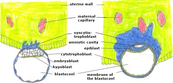
Figure 12. Implantation of the embryo in the uterine wall
Left: on day 7-8. The embryo lies against the uterine wall on the side of the embryoblast. The syncytiotrophoblast expands into the maternal tissue (yellow-green). The hypoblast is ligated
from cells of the embryoblast (white).
Right: on day 8-9. The embryo eats further into the uterine wall, the hypoblast (flat cells) has extended all the way down and forms, together with the cytotrophoblast, the membrane of the blastocoele. In the embryoblast the amnion arises. The epiblast (high cells) is located above the hypoblast and the amnion is formed from epiblast and cytotrofoblast cells. The syncytiotrophoblast lies against a maternal blood vessel (capillary).

Figure 13. Implantation, continued
Left on day 9. The embryo eats further into the uterine wall. The syncytiotrophoblast proliferates in the maternal tissue and lies around capillaries and makes holes (lacunae) where maternal blood can flow. Maternal blood and tissues remain separated from embryonic tissue. The amniotic cavity develops, by which the two-cells-thick embryonic disc arises. The tissue of the hypoblast covers the blastocoel membrane, now called yolk sac. Between the trophoblast and the membrane of the yolk sac a thick tissue develops: the extra-embryonic mesoderm.
Right on day 12. The embryo is completely enclosed by the tissue of the uterine wall. The syncytiotrophoblast is still rapidly expanding. The extra-embryonic mesoderm is thicker and holes in the extra embryonic coelom are developing.
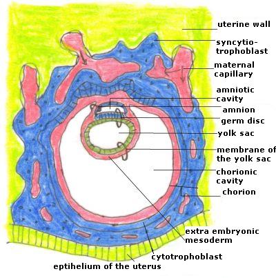
Figure 14. The embryo on day 13 (simplified)
The holes in the extra-embryonic mesoderm have joined together to form the chorionic cavity, its membrane is the chorion. The syncytiotrophoblast is growing all around the embryo, it is thicker on the inside of the uterine tissue than near the epithelium. The embryonic disc is on the backside attached to the chorion. All around the chorion cavity lies the syncytiotrophoblast with the lacunae containing maternal blood. (The primary yolk sac which is now separated from the now called secondary yolk sac has been omitted.)
Development of the embryonic disc
Flat, square cells are ligated from the embryoblast on the side of the blastocoele (Fig. 12 to 15). These cells are called the hypoblast (hypo = under). The hypoblast expands on the inside of the trophoblast, too, and covers it. The blastocoele is now called yolk sac.
In the embryoblast a small cavity develops, called the amniotic cavity, its roof is called the amnion (= sheep skin). The cells adjacent to the hypoblast become cylinder - or elongated cube-shaped; these cells form the epiblast (epi = upper).
Through the formation of the yolk sac and the amniotic cavity a round, flat embryonic - or germ disc is formed, existing of two layers (epi- and hypoblast). At the end of the week the prochordale plate (a spot of different shaped cells) develops in the embryonic disc.
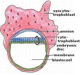
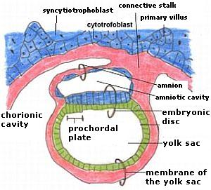
Figure 15. The embryonic disc on day 9 (left) and day 14 (right)
Left: The embryonic disc consists of two cell layers and is round and flat.
Right: at the tail-side the embryo is attached to the chorion and the cyto- and syncytiotrophoblast by the connective stalk. It hangs free in the fluid-filled chorion cavity. The prochordale plate is formed in the embryonic disc at the side where the head will develop. Epiblast cells (up) are high, hypoblast cells (under) are flat.
Origin of the chorion cavity and the connective stalk
From day 9 on, the tissue between trophoblast and embryonic disc, amnion and yolk sac thickens (Fig. 13). This tissue is called extra-embryonic mesoderm (extra = outside). A confusing name, because the tissue lies within the embryo. The name only indicates that the tissue lies outside the embryonic disc. Because the syncytiotrophoblast and the cytotrofoblast grow much faster than the germ disc, in this mesoderm tissue crevices and cavities arise (called the extra-embryonic coelom (= cavity)). From day 12 on they unite and form the chorionic cavity. The trophoblast is now called chorion (= skin or leather).
First, the germ disc with amnion and yolk sac is connected to the chorion (12 days) on the posterior side. This attachment moves to the tail (or better: the place where the tail will develop) and becomes narrower. This attachment is called the connective stalk.
The round, flat germ disc, amniotic cavity and yolk sac are attached to the connective stalk, like two halves of a ball, and they hang freely in a large spherical space (the chorionic cavity) with the chorion as a surrounding wall.
Size
The embryo grows. At the beginning of the second week its size was approximately 0.3 mm, at the end 3 - to 3.5 mm. In a one week period it grew to 10 times its size. The germ disc is still small: 0.5 mm.
Twins
About one in 90 pregnancies is a twin. Twins may grow from one or two eggs. Dizygotic twins result from two fertilized eggs, do not look the same and can either be of the same - or of different sex. They cover 2/3 of all twins. The two embryos develop as described above and both have their own amnion and chorion (Fig. 16).
Monozygotic twins are born from one fertilized egg, usually because in the first week two embryoblasts develop in the blastula. These embryos have their own amnion and share the chorion and placenta (Fig. 17).
A small number of twins develops in the second week, after the formation of the amnion, by cleavage of the embryonic disc. They are in the same amnion together and also share the same chorion and placenta (Fig. 18).

Figure 16. Dizygotic twins
There are two zygotes, which implant. They both have their own shell (amnion, chorion) and trophoblast.
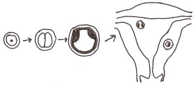
Figure 17. Monozygotic twins
There is one zygote with two embryoblasts that implant. Both embryos have their own amnion, and share the same chorion.

Figure 18. Monozygotic twins from the second week
The embryonic disc divides. The embryos are together in the same amnion and chorion.
Characteristics
In the first week the embryo developed in the shell of the zona pellucida. The single-celled zygote was divided into many small and similar cells, creating the morula and blastula. At the end, the trophoblast and the embryoblast arose. Because the embryo breaks out of the zona pellucida at the end of the week, we see a reversal in the second week: the embryo starts to grow. The cells are not getting any smaller, they can grow now. In particular, the trophoblast is growing very fast, so fast that no cell membranes are made in the syncytiotrophoblast.
The focus of growth lies at the periphery, the trophoblast. The growth of the centre, of the embryoblast, stays behind, as is shown by the tearing of the extra-embryonic mesoderm and the development of the chorionic cavity. In the centre we see a differentiation in amnion and yolk sac, in epi- and hypoblast.
There is now interaction with the environment, the isolation through the zona pellucida of the first week is gone. The embryo gives off hormones and enzymes, takes up nutrients, etc. Therefore it can grow and gain a place in the uterine wall. The shelter of the zona pellucida is replaced by the shelter of the nourishing uterine wall.
The centre, the embryonic disc, is a flat round disc of two cell layers, which has an upper and a bottom side, but no left and right. The disc has no content, as the two layers are both on the outside. An early orientation of directions occurs only at the end of the week with the formation of the connective stalk.
Monozygotic twins can develop in the first week, and at the beginning of the second week. Thereafter the embryonic disc is no longer divisible. The embryo has then become in-dividual (= not-divisible).
The development of the first week is similar in shape and time in all mammals. Now that's no longer the case, both form and duration are species-dependent.
The characteristics of this week are a reversal of the characteristics of the first week and are that of the plant:
- There is a huge growth on the outside.
- As the embryonic disc is flat and round, there is no left and right in the centre and there is no content.
Therefore, it can be said that the embryo of the second week has the characteristics of the plant and can be called the "plant-man".
Rudolf Steiner said that in the second week man is not in the embryo yet, but floats around it. The spirit lives in the periphery, in the tissue of the trophoblast.
| first week | second week | |
| development | divisions | growth and proliferation |
| communication with the environment | no | intensive |
| duration | species independent | species dependent |
| tissues | no differentiation | differentiation |
| shell | zona pellucida | uterine wall |
Table 3. Differences of the embryological processes of the first and the second week

Figure 19. The plant or a picture for the "plant-man" (from van der Wal (2003): Hartmann)
With its growth the plant is committed to the periphery; it grows from its seasonal sprout in all directions into space.