
Man does not become human,
but is always human.
Erich Blechschmidt


The fifth to eighth week: the un-folding
In the fourth week a body was formed in the centre of the embryo, clearly demarcated from the nourishing periphery.
On this page you will find:
- how the body changes until the end of the eighth week;
- the development of the limbs;
- the development of twins (as a sequel to the description of the second week);
- the development of the fetal membranes.
Changes in the embryo
In the figures 34 and 35 four side views are drawn of the embryo from the fifth to the eighth week.
On the 32nd day (Fig. 34 left) the embryo is yet more enfolded inward than at the end of the fourth week. Head and tail are bent inward and face each other. The back is round and curved. The head is large, especially the brain is large, and lies over the big heart. The limbs are like small paddles. The umbilical cord has become compact: connective stalk, yolk sac and allantois are lying close to each other.
On the 41st day (Fig. 34 right), the spine has partially stretched and the embryo is less rounded. The head has been pushed upwards by the large heart. In the umbilical cord a bump can be seen, caused by a part of the digestive tract for which there is no place in the abdomen so that it bulges out of the body. The short tail is still present. The limbs have grown and fingers and toes are becoming visible.

Figure 34. Left: the embryo on day 32, right: on day 41; 7 and 12 mm long.
On the 51st day (Fig. 35 left), the head is bent forward in relation to the unfolded back. The neck is visible as a slight curve. The head is rounder and eyes and ears are visible. The heart has been taken up into the torso, the intestines still protrude and make the abdomen big and round. Arms and legs stick out of the body and are longer. The tail has gone.
On the 56th day (Fig. 35 right), the body is a little human being. Only 3 cm long, he looks just like us. The large round head with the big forehead, eyes and ears is straight on the body. The neck has been formed. There is a convex belly, in the umbilical cord the bulging hump of the digestive tract is still visible. The hips and waist have been formed. The legs are placed under the torso.
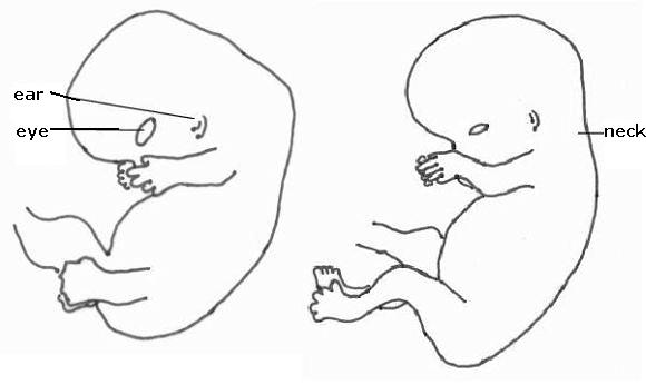
Figure 35. Left: the embryo on day 51, right: on day 56; 22 and 30 mm long.
Characteristics
In the fifth until the eighth week, the body gets its final form. First, the enfolding movement of the fourth week is continued and the embryo gets rounder. Then an unfolding movement can be seen: the spine and the back stretch and the neck and later the waist and the hips appear. Head and limbs become apart from the torso. This process goes from top to bottom: first the neck, then the waist. It is this unfolding process that makes man human. He is centred, and acts from this centre.
Neck and waist ensure that head, chest and limbs become separate areas. The threefoldness of the human body is there.
Man is the only organism that stands and goes upright: all joints (neck, shoulder, hip, knee, ankle) are in one vertical plane. When man stands, he is in balance. Standing and locomotion take little energy. Because he stands on his feet, his hands are free to use at his discretion.
Apes also go through the process of unfolding their back and the development of a neck and a waist, but they lose the erect posture again, the chimpanzee not until after birth (Verhulst).
The polarity between the closed, spherical head and the radiated, elongated limbs is visible. In many ways they are opposites with the trunk in between.
Head, trunk and limbs each have their own task: the head is for observing and thinking, the torso, with heart and lungs, for feeling and the limbs are for action.

Figure 36. Human Man (from Van der Wal, by Hartmann)
Like an animal, man is a centred organism with an inner world. The difference between them is that man not only has a physical centre, but also a spiritual centre, his self-consciousness. Man has a position from which he can look at the world and at himself and he can act freely out of his centre.
The limbs (including characteristics)
At the end of the fourth week the buds of the arms appear and two days later the buds of the legs (Fig. 33). Fig. 37 shows the development of the limbs. Of the arm the ends appear first, i.e. the bud grows into fingers. After that, the hand and then the forearm appear, followed by the upper arm and finally the shoulders. Fingers and toes appear first as flat, paddles-like forms that develop radials and after a phase in which they look like flippers, individual fingers and toes appear. On the 52nd day, the arms and fingers are fully formed, on the 56th day the legs and toes.
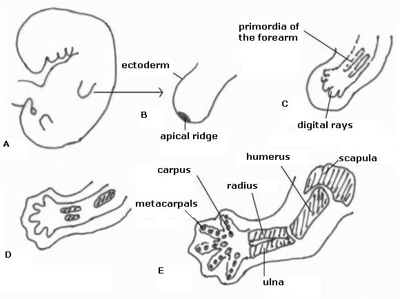
Figure 37. Development of the arm
First the bud of the hands and a day later of the
feet appear (A and B). In C the forearms are also visible, the upper arm at D, and E shows the shoulder blades too. Mesoderm slowly hardens, at first the bones are still cartilage.
The arms and legs grow lateral of the body. In the 8th week the arms turn outward (external rotation) and the legs inward (internal rotation), leaving the hands with the palms upward and the feet downward (Fig. 35). The hands are oriented to the sky, to heaven, the feet are earth-oriented.
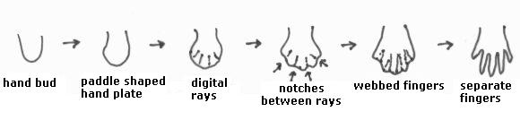
Figure 38. The development of the fingers and toes First there are paddle-shaped plates in which the fingers and toes arise like rays. In the rays, the tissue gets thicker, in between the tissue gets thinner. The thin tissue disappears and the individual fingers and toes are formed.
Fig. 35 shows that the arms lie on the heart and legs lie along the umbilical cord. The arms move with the rhythm of the embryo's own (fast) heartbeat; the legs move with the rhythm of the blood in the umbilical cord, which is determined by the heartbeat of the mother. The arms move with the rhythm of the embryo, the legs with that of the mother. The arms belong more to the embryo, the legs more to the surroundings, to the mother. From the beginning the arms are focused more on the embryo itself, the legs more on the world outside.
Man stands between heaven and earth, he is focused on the earth through his feet on the ground and focused on heaven through his hands. His feet are earthbound, his hands are free.
| arms | legs | |
| rotation | outward (external rotation) | inward (internal rotation) |
| direction | hands are up | feet are down |
| place | on the heart | along the umbilical cord |
Table 5. Differences between arms and legs
On the side of the head the embryo develops faster than on the side of the tail. The neural tube closes earlier at that side, it is at the head that the digestive tract is formed earlier; the arms develop earlier and faster than the legs. This is called the cranio-caudal (= head-tail) growth movement. This is in contrast with the limbs, where the fingers and toes appear first and the shoulders and pelvis appear last. What is closest to the centre comes last and what is furthest away comes first. This is a reversal of the cranio-caudal development movement. This second growth movement can be called the centre-periphery (or disto-proximal) growth movement (Fig. 39). This growth movement is important for humans: this long embryonic development-time, gives more time to develop what arises late. As a consequence, the legs grow longer than the arms and the upper legs longer than the under legs (Verhulst). This growth movement largely determines the upright posture of man.

Figure 39. Left: cranio-caudal growth movement
and right: centre-periphery growth movement
(Verhulst)
Twins
In addition to what has been said in "The second week" and shown in Fig. 5, Fig. 40 shows drawings of the three possible types of twins. From left to right:
- Both embryos of non-identical twins each grow in their own chorion and amnion, the placentas can be separated or connated.
- The fraternal twins of the first week developed from two embryoblasts within one trophoblast. The embryos share the chorion and the placenta and each grow in their own amnion.
- The fraternal twins of the second week arose from a division of the embryonic disc. These embryos share amnion, chorion and placenta. There is considerable danger of the umbilical cords winding around each other, which may cause that the blood supply of either to be cut off.
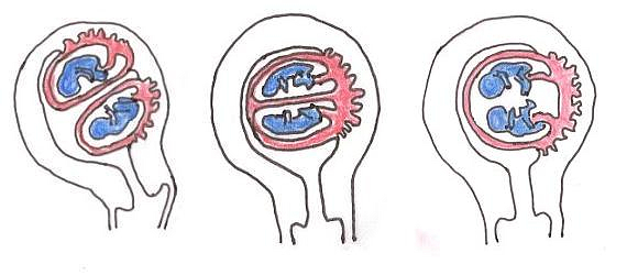
Figure 40. Twins, from left to right: non-identical twins, fraternal twins in the first week, fraternal twins in the second week. Explanation in the text.
The fetal membranes
The embryo is - from inside to outside - enveloped in the amnion, the chorion and the uterine wall. The embryo grows and is called a fetus after the eighth week. In the drawing of an embryo of the fourth week in Fig. 41A, the amnion is still small and the embryonic disc with the amnion and yolk sac hangs freely in the chorionic cavity. In Fig. 41B, the amniotic cavity has grown and fills almost the whole space of the chorionic cavity. The uterine cavity is almost entirely occupied by both. Fig. 41C. shows that the uterine cavity is entirely occupied by the fetus. The chorionic cavity is almost entirely filled by the amniotic cavity. Eventually, chorion and amnion coalesce (Fig. 41D). The embryo thus floats in amniotic fluid.
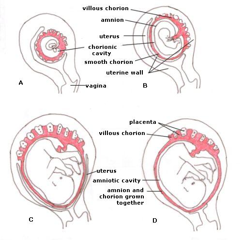
Figure 41. The development of the fetal membranes and the placenta. A: 4th week, B: 8th week, C: 16th week, D: 22nd week. See text for a description.
Images of man
When is the embryo human? Is it always human, or is it only human from a certain point or a certain appearance? An embryo of 8 weeks certainly looks human. A single-cell zygote is more like other single-celled organisms and does not look like a human. The embryonic stages of the fourth week look strange and not very human.
An answer can be found by looking back at the embryonic development. An adult looks different from a teenager and behaves differently. A teenager looks different from a child and a child looks different from a baby. There is a continuous line to be seen and although a child and a baby look different from- and behave differently than adults, we see them as a young humans.
What about the fetus? It looks like a baby but shows different behaviour in a different environment. When a baby is born prematurely, every effort is made to keep it alive.
The fetus developed from the embryo, which developed from the embryonic disc, which developed from the blastula and finally from the zygote. All are manifestations of a human being. All manifestations have their own appearance and behaviour, appropriate for their stage in life and the environment they are in. So, just as the baby differs from the adult, the zygote is different from the baby. This leads to my conclusion that the zygote and the embryo are human, in a different form. Hence: embryology: images of man.